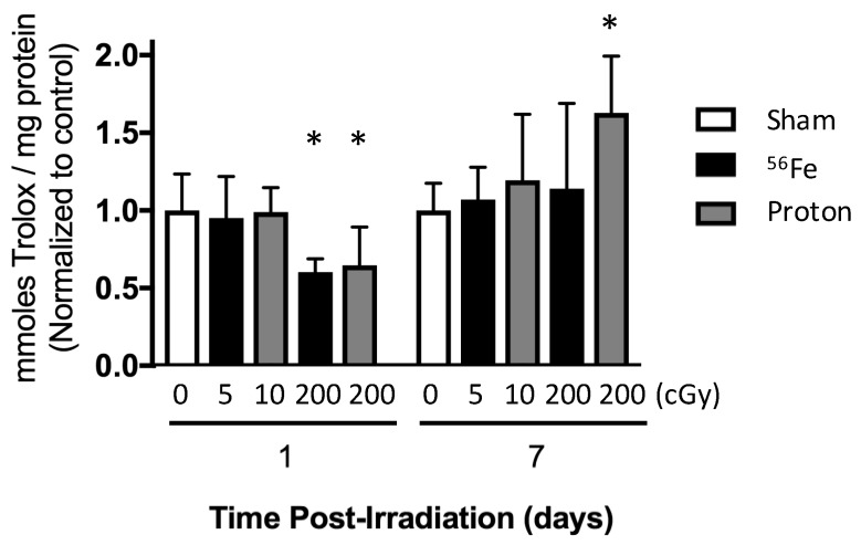
Figure 6.
Irradiation perturbation of the marrow oxidative milieu, assessed by the antioxidant capacity of the extracellular fluid (ECF), in mice at one and seven days post exposure (following timeline in Figure 1B). Mice were irradiated with 56Fe at 5 or 200 cGy or protons at 10 or 200 cGy. Data are mean ± standard deviation. * p ≤ 0.05 versus sham control.

