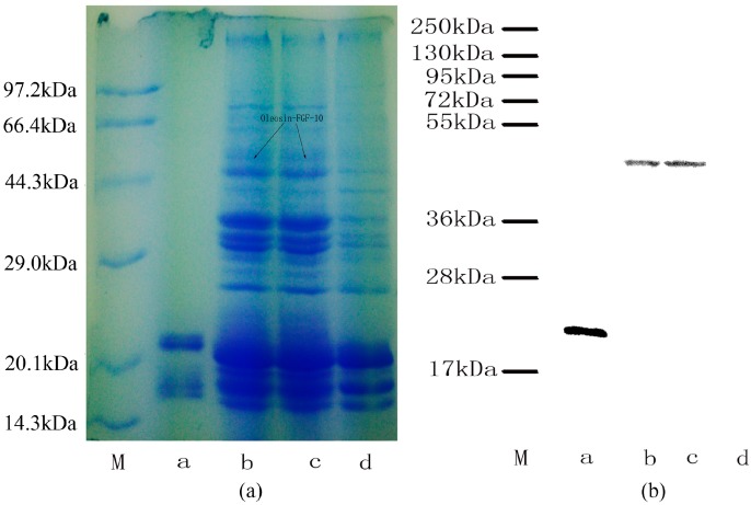
Figure 2.
Protein analysis of oleosin-rhFGF-10. (a) Oil body proteins after 12% denaturing gel electrophoresis and Coomassie brilliant blue R250 staining. Lane M, molecular weight standards, lane a, rhFGF-10; lanes b–c, the same transgenic safflower lines; lane d, wild-type safflowers. Black arrows indicate the protein of interest and its molecular weight of 45 kDa; (b) western blot detection of the oleosin-rhFGF-10 protein. Lanes are as for (a).

