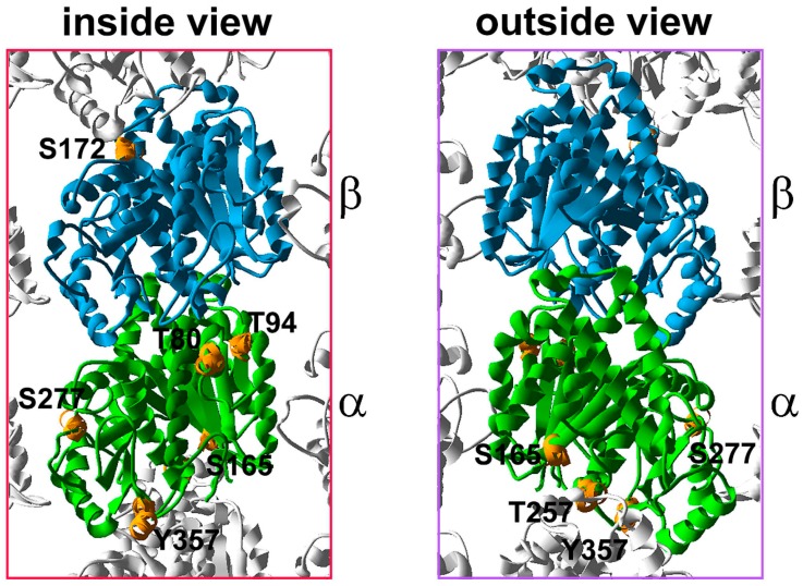
Figure 3.
The distribution of the phosphorylated residues within the α,β-tubulin heterodimer. The 3D model of the tubulin heterodimer within the microtubule lattice is based on 3J6F.pdb (NCBI database), an inside and outside view. Putative and confirmed sites of tubulin phosphorylation are marked in orange. Luminal T80 and T94 of α-tubulin are located within the H2 helix and S3 strand, respectively. α-Tubulin S165 is located within the H4–S5 loop, near the H3 surface, but is not a part of this surface. α-Tubulin T257 is located within the H8 helix, building the minus end surface. α-Tubulin S277 is located in the S7–H9 loop, also called the M loop but according to the model proposed by Inclán and Nogales [9] S277 is not a part of the ML surface. A modification of S277 could influence the structure of the ML surface and thus affect the interactions with the neighboring protofilament. α-Tubulin Y357 is located within the S9–S10 loop. S172 of β-tubulin is located within the S5–H5 loop, called also T5 loop.

