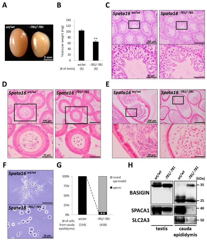
Figure 4.
Spermiogenic arrest in Spata16−781/−781 mice. (A) Testes in wild-type and Spata16−781/−781 mice. Scale bar: 2 mm; (B) Testicular weights in wild-type and Spata16−781/−781 mice. Testicular weights in Spata16−781/−781 mice were significantly reduced compared with those in wild-type mice. ** p < 0.005; (C) PAS staining of testicular sections. Lower figures are magnified images of the boxes indicated in the upper figures. Scale bars: upper; 200 μm and lower; 50 μm; (D) HE stained sections of caput epididymis. Lower figures are magnified images of the boxes indicated in the upper figures. Scale bars: upper; 100 μm and lower; 50 μm; (E) HE stained sections of cauda epididymis. Lower figures are magnified images of the boxes indicated in the upper figures. Scale bars: upper; 200 μm and lower; 50 μm; (F) Observation of cells extracted from cauda epididymis. Scale bars: 50 μm; (G) Quantitative morphometric analysis of round spermatid and sperm from cauda epididymis. Dashed line indicates a significant reduction in sperm-like cells extracted from the cauda epididymis in Spata16−781/−781 mice. ** p < 0.005; (H) Immunoblot analysis of cell lysates collected from a testis and cauda epididymis. Sperm acrosomal protein SPACA1 remained in the Spata16−781/−781 mouse testis. In cauda epididymal lysates from Spata16−781/−781 mice, the aberrant retention of testicular BASIGIN was detected and sperm tail-specific protein SLC2A3 and SPACA1 were decreased. Spata16−781/−781 male mice were sterile because of a spermiogenic arrest. 20 μg of cell lysates were loaded in each lane.

