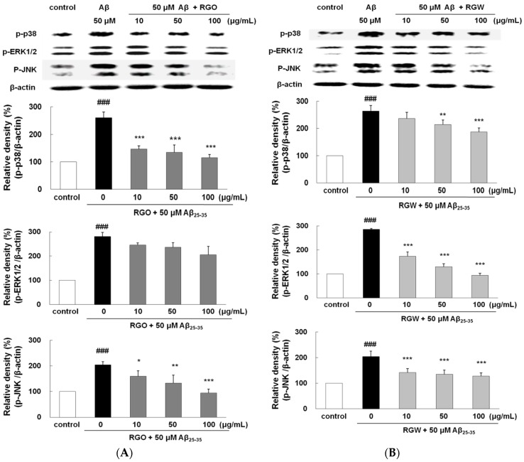
Figure 8.
RGO (A) and RGW (B) ameliorated Aβ25–35-stimulated MAPKs phosphorylation. PC12 cells were treated with Aβ25–35 for 1 h and then cellular lysates were analyzed by Western blot assay. The separated proteins were analyzed by Western blot analysis as described in Materials and Methods. ### p < 0.001 compared to control group; *** p < 0.001, ** p < 0.01 and * p < 0.05 compared to Aβ25–35 alone (ANOVA followed by Duncan’s multiple comparison test).

