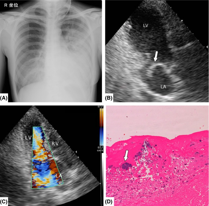
Figure 2.

Imaging and histological findings of Case 2, a 40‐year‐old woman with unilateral pulmonary edema. A, Anteroposterior chest radiograph shows left‐side limited infiltrates without cardiomegaly. B, Transthoracic echocardiography shows mitral valve prolapse (arrow). C, Color Doppler transthoracic echocardiography reveals mitral valve prolapse with severe regurgitation and the regurgitant jet tending to blow leftward within the left atrium. D, Histological findings reveal inflammatory cells and fibrin deposition in the mitral valve, and vegetation with Gram‐positive cocci infiltration (arrow). LA, left atrium; LV, left ventricle; RV, right ventricle.
