
Figure 1.
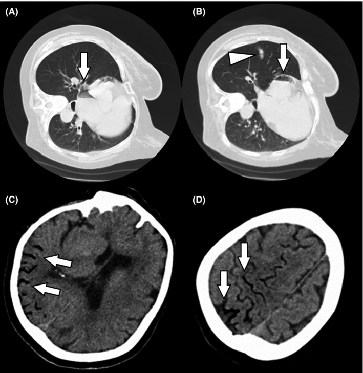
Computed tomography (CT) findings obtained at clinical onset of cerebral air embolism in a 74‐year‐old woman who underwent computed tomography‐guided transthoracic needle biopsy of a small lung mass. A, B, Chest CT showing small pulmonary hemorrhage (arrowhead) and air in the right and left atria (arrows). C, D, Brain CT showing air inflow in the right middle cerebral artery (arrows).