
Fig. 4.
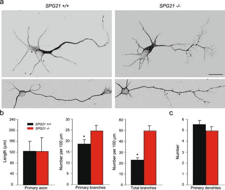
Cerebral cortical neurons from SPG21−/− mice exhibit increased axon branching. a Representative neurons at 3 days in vitro were co-stained for β-tubulin and tau to identify neuronal processes. Scale bar, 20 μm. b, c Quantifications of primary axon length and number of total axon branches (b) as well as the number of dendrites per cell (c) are shown graphically (means ± S.D.; *p<0.05)