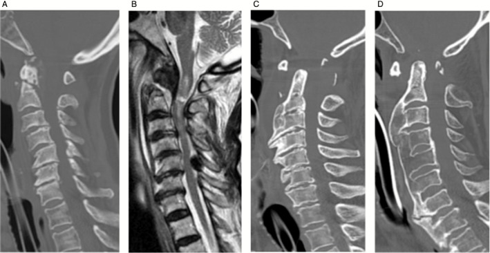
Figure 2.

(A) Computed tomography (CT) scan shows atlanto‐odontoid joint degenerative change and type 2 odontoid fracture in case 3. (B) Magnetic resonance imaging (MRI) shows upper cervical laceration in case 3. (C) CT scans show vertical type atlanto‐axial dislocation associated with DISH in case 8. (D) CT scans show lateral type atlanto‐axial dislocation associated with diffuse idiopathic skeletal hyperostosis (DISH) in case 9. OPLL, ossification of the posterior longitudinal ligament; SCI, spinal cord injury.
