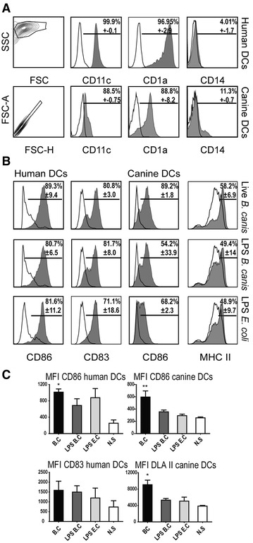
Figure 2.

DC cultures purity and activation. DC differentiation from blood purified monocytes in the presence of GM-CSF and IL-4 was identified by flow cytometry according to forward scatter (FSC) and side scatter (SSC) parameters. Gated human and canine DCs were analyzed for expression of CD1a, CD11c, and CD14 and B. canis-stimulated labelled DCs (shaded histograms) were compared to FMO (fluorescence minus one) control (black solid line) (A). Activation and maturation levels of DCs after stimulation with B. canis: human and canine DCs were identified according to FSC and SSC parameters, and gated cells were analyzed for expression of CD86, CD83, and DLA II (shaded histograms) and compared to non-stimulated cells (black solid line) (B). Mean fluorescence intensity (MFI) for each surface marker shown in B (C). *p < 0.05 and **p < 0.01.
