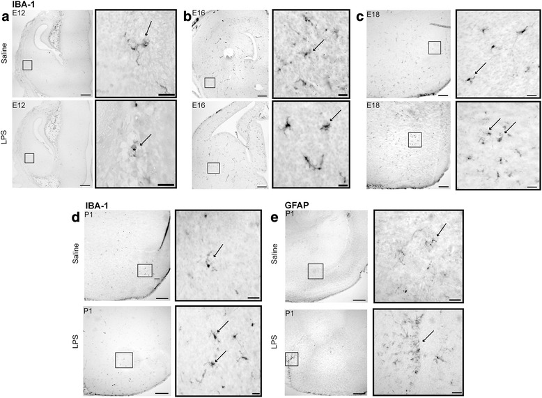
Fig. 3.

MIA induced changes in microglial morphology in the developing amygdala. Iba-1 immunoreactivity (ir) at a E12, b E16, c E18, and d P1. GFAP expression was first present at P0 with no GFAP expression preceding this time point (e). Iba-1 positively labeled microglia exhibited ramified and amoeboid morphologies from E12 to P0. In LPS-treated groups, microglial morphology was predominantly amoeboid with GFAP immunoreactivity being dense along the external capsule of the amygdala. Astrocytes were hypertrophic in the LPS-treated group at P1. Scale bar = 200 μm and inset images scale bar = 50 μm
