Figure 1.
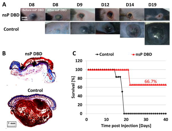
The nsP DBD treatment eradicates the melanoma tumor. (A) B16 Melanoma cells were injected on the rear flank of C57BL/6 mice. After 8 d, the tumor was treated a single time for 7 min with nsP DBD at 236 Hz and 33.6 kV. The control tumor continued to grow up to day 19 (D19) post-injection until it was sacrificed. Mice treated with nsP DBD continued to heal with a scab up to 22 days and after were tumor free. (B) Trichrome staining of D22 nsP DBD treated tumor (top) and Control tumor (bottom) on D19 post injection. Histology of the nsP DBD treated tumor shows red skin staining confirming scab formation but no tumor below the epithelium is visible. (C) Survival for nsP DBD treated tumors (red triangle) and control untreated tumors (black diamond) as a function of time post-injection (n = 6 each). The nsP DBD treatment resulted in significant improvement of survival rate (66.7% post-treatment) compared to control (0%, p <0.005).

