Figure 2.
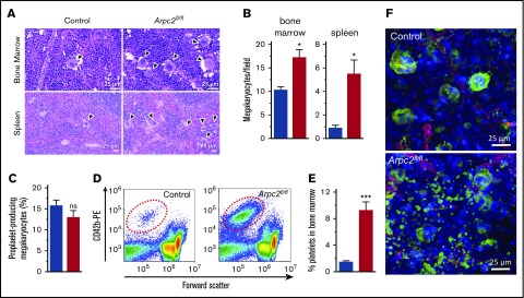
Premature platelet release into the bone marrow compartment in Arpc2fl/flPF4-Cre mice. (A) Increased megakaryocyte density in representative images of bone marrow and spleen sections stained with hematoxylin and eosin. (B) Quantification of megakaryocyte density in indicated organs (8-13 random fields analyzed per organ; n = 3). (C) Percentage of proplatelet forming bone marrow megakaryocytes observed in culture (n = 7-8). (D) Representative flow cytometry dot plots showing increased number of platelet-sized CD42b+ particles in bone marrow from Arpc2fl/flPF4-Cre mice. (E) Quantification of CD42b+ particles in bone marrow (n = 6). (F) Representative confocal fluorescence images of bone marrow from the indicated mice. Green, megakaryocytes/platelets; red, endothelial cells/vasculature; blue, nuclei; blue bars, control; red bars, Arpc2fl/flPF4-Cre. Data presented as mean ± SEM, *P < .05, ***P < .001.

