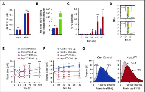
Figure 4.
Loss of Arp2/3 leads to macrophage-mediated clearance of platelets. (A) Binding of RCA-fluorescein isothiocyanate to platelets from mice treated (+neu) or not (−neu) with neuraminidase (n = 4-6). (B) Detection of platelet-associated antibodies with anti-mouse–IgG-488 (green bar, positive control). (C) Kinetics of phosphatidyl serine exposure in washed platelets incubated in the presence of 4 mM CaCl2 (n = 6). (D) Representative flow cytometry plot illustrating the interaction of RAW 264.7 macrophages and fluorescently labeled platelets. Changes in peripheral platelet count (E) and platelet size (F) in indicated mice following macrophage depletion by clodronate liposomes (lip) or treatment with PBS liposomes (n = 10). (G) Frequency analysis of circulating platelet size 72 hours after treatment with clodronate liposomes or PBS liposomes (mean distributions of 5-6 mice per group). Blue bars/lines, control; red bars/lines, Arpc2fl/flPF4-Cre. Data presented as mean ± standard deviation. *P < .05, ***P < .001. FSC-H, forward scatter height.

