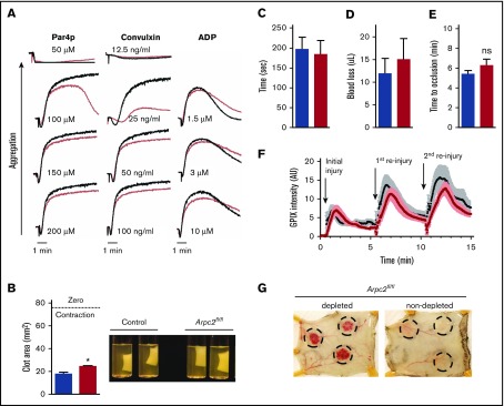
Figure 6.
Mild platelet activation defect but intact hemostasis in Arpc2fl/flPF4-Cre mice. (A) Representative traces showing aggregation of platelet rich plasma stimulated with increasing doses of the indicated agonists. (B) Quantification of total clot area after platelet-mediated retraction (n = 3-5) and representative images of retracted clots. (C-D) Tail clip bleeding assay. Time to cessation of bleeding (C) and amount of blood loss (D) from severed tails of the indicated mice (n = 11-12). (E) Time to occlusion of carotid artery after induction of thrombosis by administration of FeCl3 solution to vessel (n = 8-9). (F) Kinetics of platelet accumulation and peak platelet density at injury site of saphenous vein after laser ablation (8 total injury sites per group; 3 mice per group). (G) Representative images of intradermal bleeding at sites of local inflammation induced by rPA reaction in platelet depleted and nondepleted Arpc2fl/flPF4-Cre mice (n = 3 mice). Blue/black bars/lines, control; red bars/lines, Arpc2fl/flPF4-Cre). Data presented as mean ± SEM. *P < .05.

