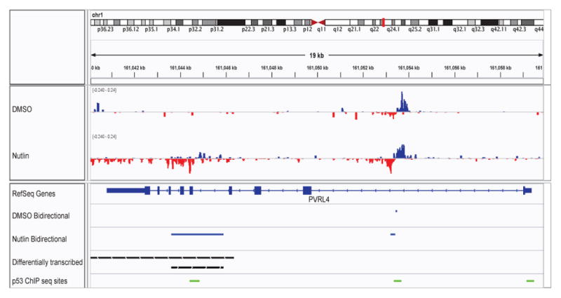
Fig. 12. Differential Transcription at PVPR4.

An IGV snapshot showing PVPR4, a negative strand gene where a small portion of the gene is differentially transcribed. The region of differential transcription (black bars) overlaps both FStitch bidirectional calls (blue bars) and p53 binding sites (green bars), indicating this may be an intragenic enhancer. The tracks, in order, are: histograms of the GRO-seq signal observed in DMSO and Nutlin, respectively (positive strand: blue; negative strand: red); RefSeq annotation for PVPR4; FStitch bidirectional calls in both DMSO and Nutlin, respectively (blue bars); FStitch differential transcription calls (black bars: top is negative strand, bottom is positive strand); location of p53 binding events (in green).
