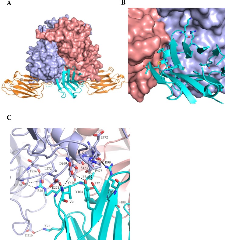
Fig 7. Nano-26 Nano-85 GII.10 P domain double complex structure.
GII.10 P domain Nano-26 Nano-85 crystal diffracted to 2.3Å in a C121 space group. Unit cell contained a P domain dimer with two Nano-85 and two Nano-26 molecules. (A) GII.10 P domain is colored as in Fig 3 with Nano-26 (cyan) and Nano-85 (orange). The Nano-85 and Nano-26 binding site in a double complex were identical to binding sites in individual complexes. (B) Nano-26 binds to the cleft between two P domain monomers at the bottom of the P domain dimer. (C) Close up view on the interactions between P domain residues and a Nano-26. Seven direct hydrogen bonds formed between P domain chain B: D269, L272, E274, E471, E472, T276 and Nano-26: V2, R26, R99, and Y104. P domain chain A: I231, P488 and chain B: E271, D316, Y470, and P475 were involved in hydrophobic interactions and two electrostatic interactions with Nano-26: V2, I28, F30, M31, K75, and A102.

