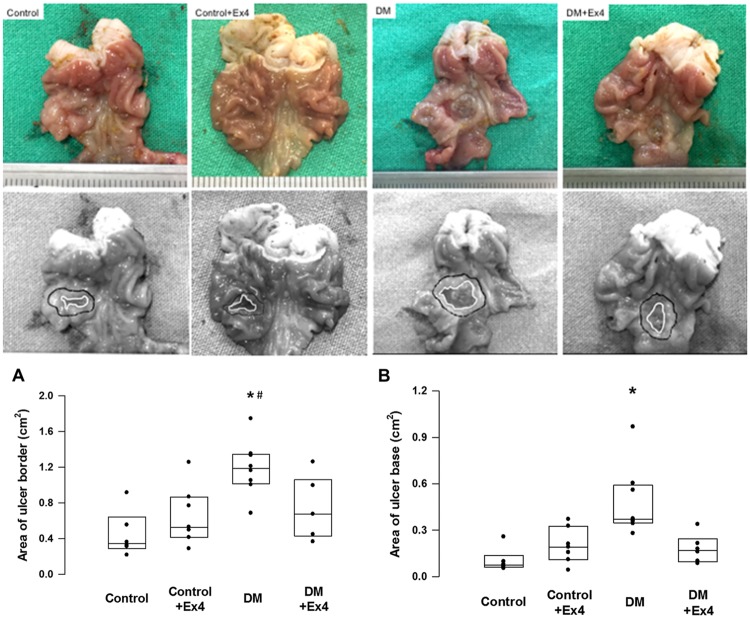
Fig 1. Representative photographies of gastric ulcer in controls and diabetic rats.
Ulcer areas were determined by measuring the margins of mucosa (ulcer border) and ulcer base at day 7 after creation of gastric ulcer. Both ulcer border and ulcer base areas were significantly enlarged in diabetic rats, and were potentitated by exendin-4 (Ex4) treatment. The ulcer areas were quantified using the ImageJ Software. Data were analyzed by the one-way ANOVA, and are presented as median (interquartile range). *P< 0.05 DM vs control, #P<0.05 DM vs DM+Ex4. n = 8–10 different animals in each group.

