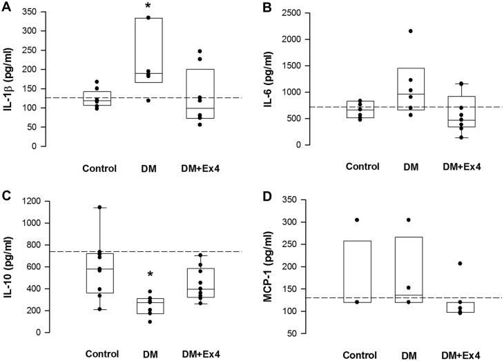
Fig 2. Quantification of tissue concentrations of inflammatory cytokines in the peri-ulcer gastric tissues at day 7 after creation of gastric ulcer in controls, diabetic (DM) rats and diabetic rats treated by exendin-4 (DM+Ex4) using ELISA kits.
Concentrations of interleukin (IL)-1β, IL-6, IL-10 and monocyte chemotactic protein (MCP)-1 were analyzed. Data were analyzed by the one-way ANOVA, and are presented as median (interquartile range). *P< 0.05 DM vs DM+Ex4 in IL-1β, and DM vs control in IL-10. n = 4–9 different animals in each group. Dashed lines indicate the mean concentrations of sham-operated animals.

