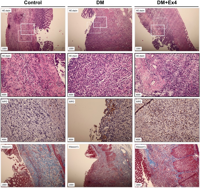
Fig 5. Representative histological sections of gastric ulcers in the three treatment groups at day 7 after creation of gastric ulcer.
The upper two panels are tissue stained with hematoxylin & eosin (HE) stain, the third panel is immunohistochemical staining for myeloperoxidase, and the forth panel is tissue stained with Masson’s trichrome stain. Solid-line boxes indicate the magnified view (400x) of HE stained tisses, in which more polymorphonuclear leukocyte infiltration. Dotted-line boxes highlight the immunostaining of myeloperoxidase (arrows) in the peri-ulcer gastric tissue. The Masson’ trichrome stain shows fragmented and disorganized collagen fibers in the peri-ulcer gastric tissue of diabetic rats. Histological sections were performed in 6 different animals in each group.

