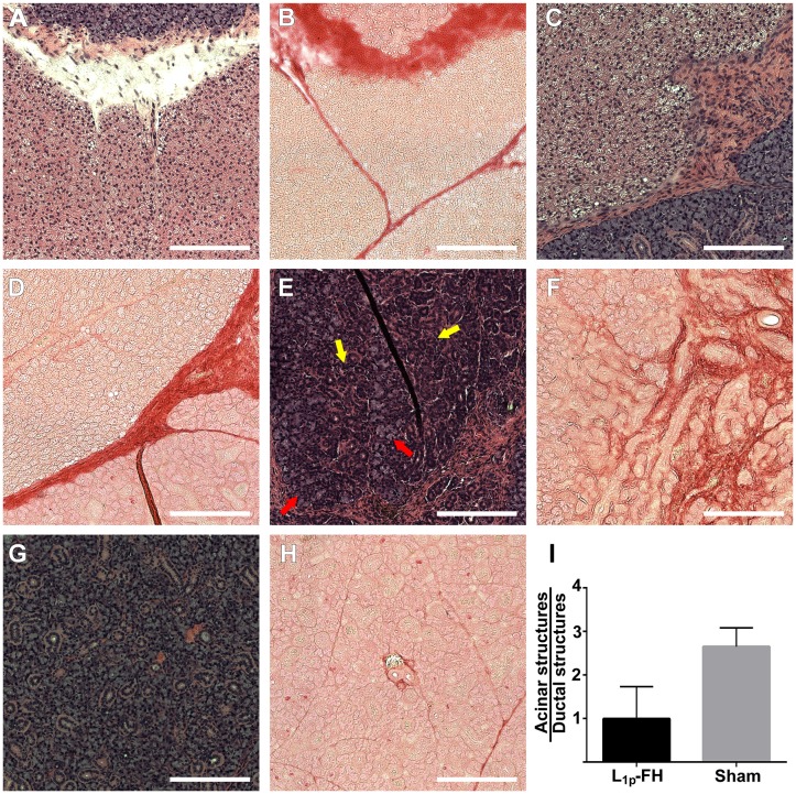
Fig 6. Surgical wounds treated with L1p-FH displayed organized mSMG.
Rehydrated sections were stained with hematoxylin-eosin (A, C, E, G) or picrosirius red (B, D, F, H) stains and analyzed using a Leica DMI6000B at 10× magnifications. Shown are wounded mSMG without scaffold (A, B), wounded mSMG with FH alone (C, D), wounded mSMG with L1p-FH (E, F), and sham control (G, H). (I) The ratio of acinar and ductal structures was analyzed using ImageJ. Red arrows indicate acinar structures and yellow arrows indicate ductal structures. Scale bars = 200 μm.

