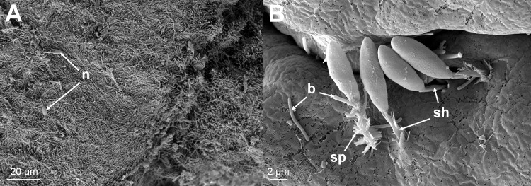
Fig 11. SEM micrographs.
(A) A SEM micrograph from the margin of a lesion showing a mat of Tenacibaculum-like bacteria with a few attached nematocysts (n). (B) A higher magnification of one of these areas showing the nematocysts with their spiked (sp) covered shafts (sh) embedded in the skin. Bacteria (b) can be seen in close proximity with some seeming to be entering the holes created by the nematocysts.

