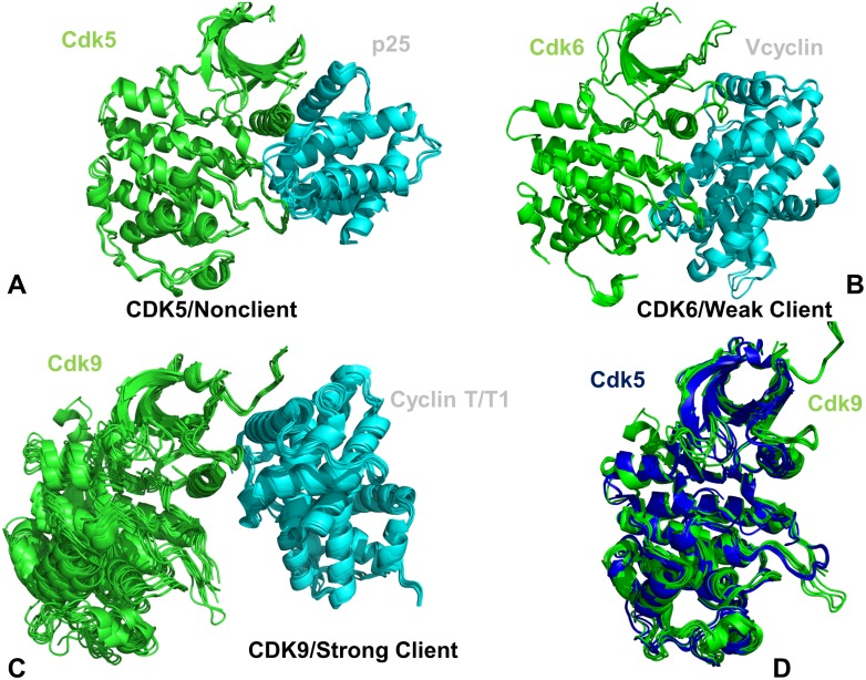
Fig 1. Crystal structures of the CDK5-p25, CDK6/V-cyclin and CDK9-cyclin T complexes.
(A) The crystal structures of the panel of CDK5-p25 complexes (pdb id 14H4L, 1UNG, 1UNH, 1UNL, 3O0G, 4AU8). (B) The crystal structures of CDK6/V-cyclin complexes (pdb id 2EUF, 2F2C) and CDK6 complexes withy inhibitors (pdb id 5L2I, 5L2S, and 5L2T). (C) The crystal structures of CDK9-cyclin T/T1 complexes (pdb id 3BLH, 3BLQ, 3BLR, 3LQ5, 3MIA, 3TN8, 3TNH, 4BCF, 4BCH, 4BCI, 4BCJ, 4EC8, 4EC9, 4IMY). The catalytic domains in panels (A)-(C) are shown in green ribbons, cyclins in cyan ribbons. (D) The superposition of the catalytic domains from the CDK5 structures (blue ribbons) and CDK9 structures (green ribbons).

