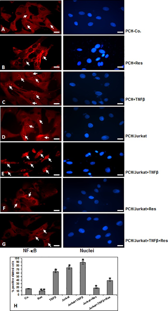
Fig 2. Immunohistochemical analysis of p65 localization after treatment with TNF-β and/or T-lymphocytes and/or resveratrol as revealed by immunofluorescence microscopy in chondrocytes.

A-G: NF-κB-p65 localization in human chondrocytes after treatment with resveratrol, TNF-β and/or co-culture with T-lymphocytes as described in Material and Methods. For immunolabeling, cells were incubated with primary antibodies against NF-κB-p65, followed by incubation with rhodamine-coupled secondary antibodies and counterstaining with DAPI to visualize cell nuclei. Original magnification, x400; bar, 30nm. To quantify the chondrocytes with nuclear-positive translocation p65 (H), the labeled cultures were examined by counting 100 cells from 10 microscopic fields. The results are provided as the mean values with S.D. from three independent experiments. Values were compared with the control, and statistically significant values with p<0.05 are designated by (*), and p<0.01 is designated by (**).
