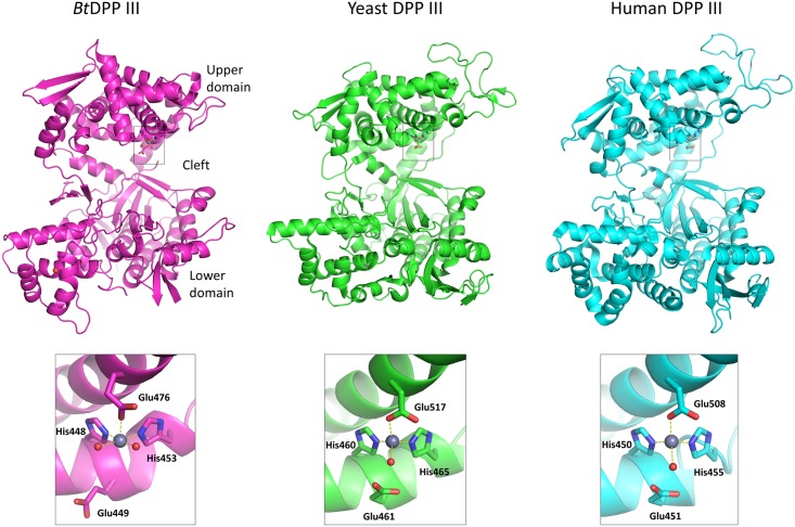
Fig 2. Structures of ligand-free Bt, yeast and human DPP III with their respective zinc binding sites.
Zinc binding sites are shown in grey squares. Amino acids coordinating the zinc ions (shown as grey spheres) and the glutamic acid residues essential for enzyme activity are shown in stick representations. The figure was prepared using the PyMol program (http://www.pymol.org/), and the PDB-deposited crystal structures of the yeast (PDB ID 3csk) and human DPP III (PDB ID 3fvy).

