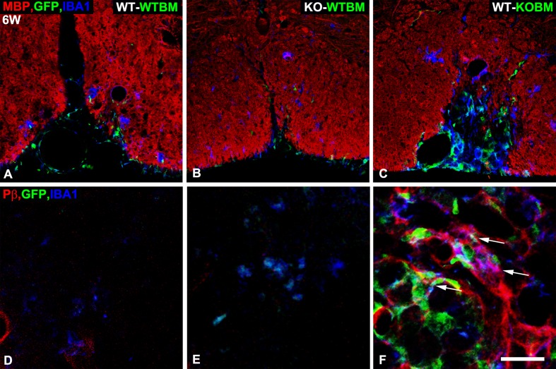
Fig 2. Persistence of atypical NG2 null macrophages in lesions in WT-KOBM mice.
Sections from lesions at 6-weeks after lysolecithin injection were evaluated for myeloid cell abundance by use of the EGFP marker (green) and immunolabeling for Iba-1 (blue). A-C. Immunolabeling for MBP (red) allows visualization of the extent of myelin repair. A. WT-WTBM. B. KO-WTBM. C. WT-KOBM. IBa-1-positive, EGFP-positive macrophages remain prominent in areas of the WT-KOBM spinal cord in which myelin repair is incomplete. D-F. Immunolabeling for Iba-1 and PDGFRβ (red) in conjunction with the EGFP marker identifies atypical macrophages that express PDGFRβ. D. WT-WTBM. E. KO-WTBM. F. WT-KOBM. Many persistent macrophages in lesions in WT-KOBM mice co-express Iba-1, EGFP, and PDGFRβ (arrows). Scale bar = 100 μm (A-C) and 25 μm (D-F).

