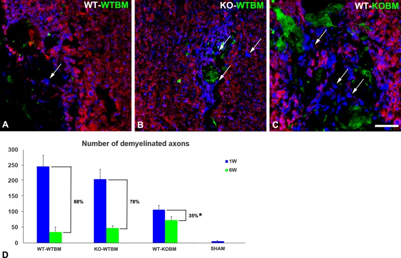
Fig 5. Efficiency of myelin repair at 6 weeks after demyelination event.
Double immunostaining for neurofilament protein (NF; blue) and myelin basic protein (MBP; red) was used to quantify numbers of demyelinated axons at 1 week (blue bars in panel D) and at 6 weeks (green bars in panel D) after the demyelinating event. Images in panels A-C are representative of the situation at the 6-week time point. Unmyelinated axons (arrows) are most numerous at the 6-week time point in lesions in WT-KOBM mice. Numbers of unmyelinated axons are determined per 0.1 mm2 of lesion area. % remyelination efficiency values (brackets) are determined after subtracting the number of unmyelinated axons (5) seen in sham operated control animals from each of the experimental values. Scale bar = 30 μm. * p < 0.05.

