
Extended Data Figure 8. Relative compound sensitivity of epithelial versus mesenchymal state cancer models.
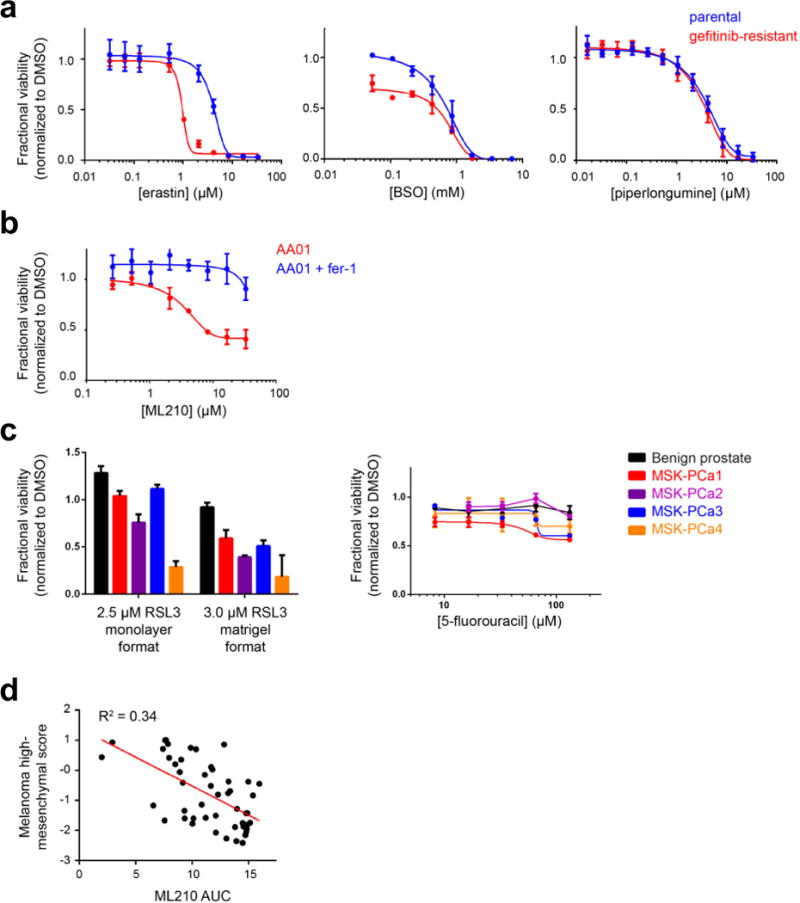
a, HCC4006 lung cancer cells that have undergone EMT as a mechanism of resistance to gefitinib (red curve). Erastin and buthionine sulfoximine (BSO) are ferroptosis inducers, while piperlongumine is an electrophile that induces a non-ferroptotic form of oxidative cell death. b, Mesenchymal state patient-derived pancreatic cancer cells (AA01) undergo ferroptosis in response to GPX4 inhibition as evidenced by the ability of ferrostatin-1 to rescue loss of viability due to GPX4 inhibition. c, Patient-derived prostate cancer organoid lines show sensitivity to a GPX4 inhibitor (RSL3) in a manner correlated with mesenchymal gene-expression score (Fig. 3d), in both collagen-based and Matrigel-based culture conditions. This correlation with mesenchymal score is not seen for a control lethal agent (5-fluorouracil). d, Scatter plot with linear regression line (red) showing the correlation between a melanoma-specific high mesenchymal state score and sensitivity to a GPX4 inhibitor (ML210) across 49 melanoma-derived cell lines from CTRP Data plotted in a–c are mean ± s.d. of four technical replicates (a and b) and three technical replicates (c) and are representative of two biological replicates.