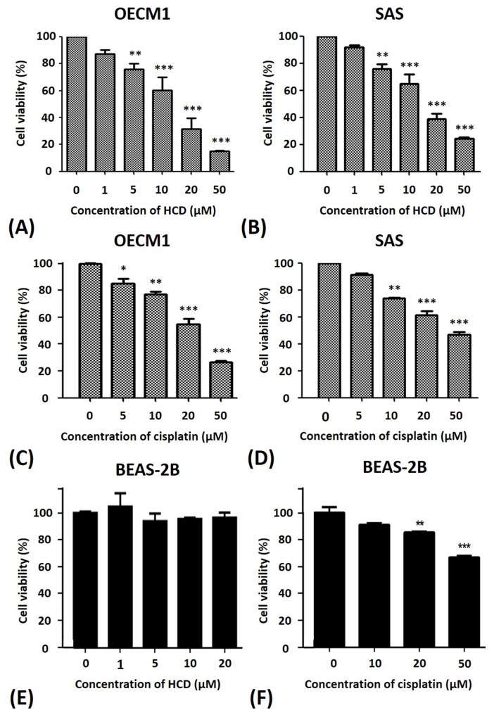
Figure 1. Alterations of the cell viability of OECM1 and SAS cells after HCD and cisplatin treatments.
(A) OECM1, (B) SAS, and (E) BEAS-2Bcells were incubated with 0 to 50 μM of HCD for 24 h, and the cell viability was determined by a MTT assay. Cell viability of (C) OECM1, (D) SAS, and (F) BEAS-2Bafter cisplatin treatment for 24 h are also shown. The data are presented as the mean ± SE of three independent experiments. * P < 0.05, ** P < 0.01 and *** P < 0.001 when compared with the untreated control (0 μM).

