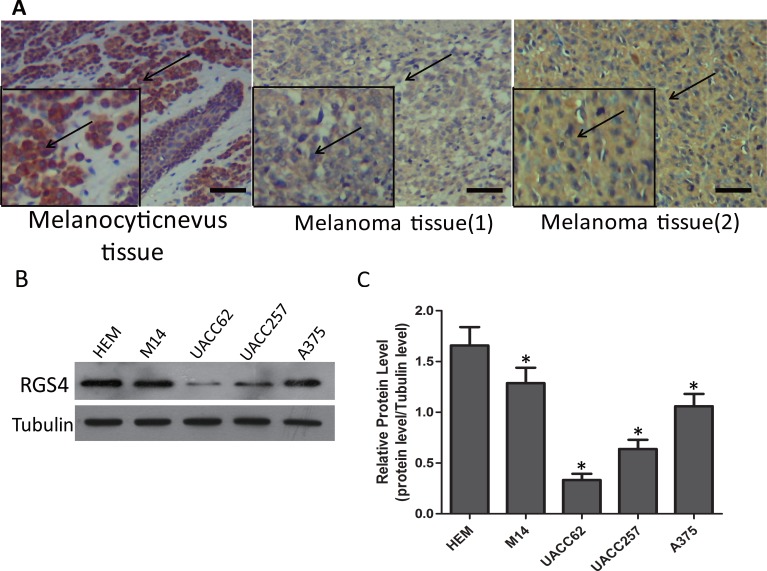
Figure 1. RGS4 expression is remarkably decreased in melanoma tissues and cells.
(A) Immunohistochemistry staining of RGS4 in melanocytic nevus tissues and melonoma tissues with anti-RGS4 antibody . Scale bars: 100 μm. (B) Western blot to show the protein levels of RGS4 varies in different normal skin cell lines and melanoma cell lines. (C) Quantification assay of the RGS4 bands intensity. Error bars indicate ± SD. *p < 0.05; **p < 0.01 by Student’s t-test.

