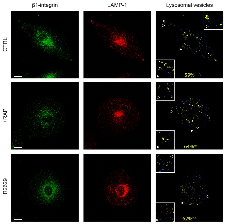
Figure 7. LRP-1 does not address β1-integrin to lysosomes.
FTC-133 cells were plated onto 1% gelatin coated coverslides and then maintained during 1 h under control conditions (CTRL), 500 nM RAP (+RAP) or 2.5 μg.mL-1 R2629 antibody (+R2629) treatment. β1-integrin staining with Alexa Fluor 488 (left panel) and infection with lysotracker for LAMP-1 (middle panel) were carried out before confocal microscopy analysis. On the right panel, lysosomes containing no β1-integrin are represented in blue and β1-integrin-containing lysosomes are represented in yellow. The average percentage of lysosomes positive for β1-integrin is indicated in yellow. Statistical analysis was conducted on lysosomal vesicles from 60 to 200 voxels (n.s., not significant; as compared to CTRL). Cell areas indicated by full and empty white arrowheads highlighting representative lysosomes for each condition are proposed as 2-fold enlargement insets. Bars: 10 μm.

