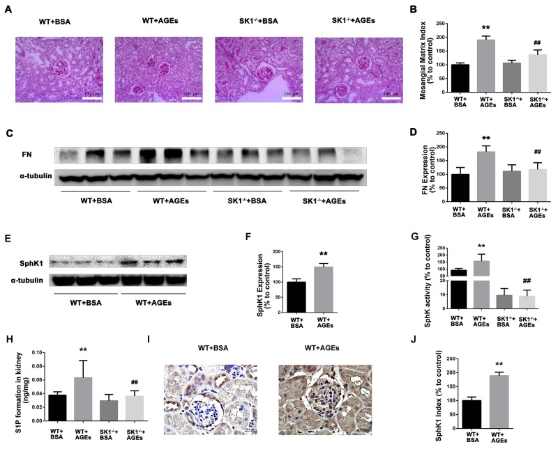
Figure 6. Effects of knock out SphK1 on FN and SphK-S1P signaling pathway in AGEs-induced mice kidney.
The pathological analysis of glomerular was performed using PAS staining (A). Original magnification, ×400. Mesangial matrix index was defined as the PAS-positive area (B). The expression of FN was evaluated by western blotting (C). The densitometry analysis demonstrated that renal protein levels of FN were increased in untreated diabetic mice and reduced by SphK1 knockout (D). The expression of SphK1 was evaluated by western blotting (E). The densitometry analysis demonstrated that renal protein levels of FN were increased in AGEs-induced mice (F). Kidney lysates were subjected to SphK activity assay (G), and Sphingosine 1-phosphate (S1P) quantification (H) using LC-MS/MS. Immunostaining showed SphK1 (dark brown) expression in glomeruli was increased at 12 weeks after AGEs induction (I). The staining of SphK1 was quantified (J). **P < 0.01 vs. WT+BSA, ## P < 0.01 vs. WT+AGEs.

