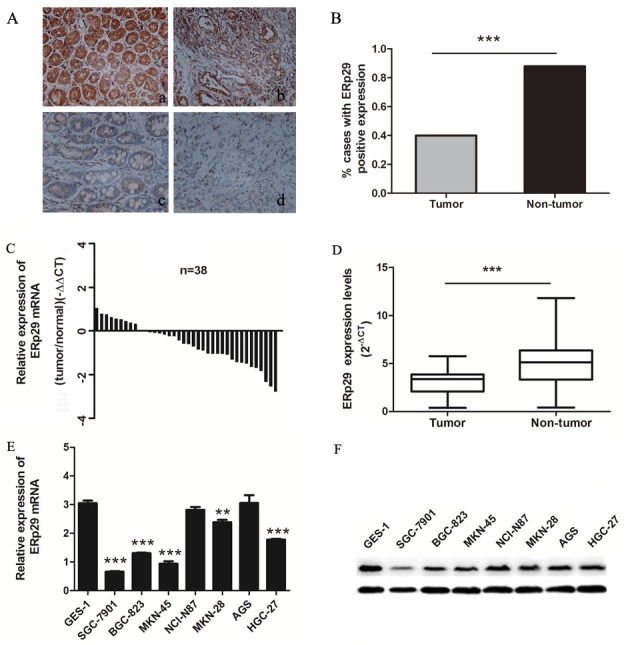
Figure 1. Expression of ERp29 in gastric cancer tissues and cell lines.

(A) Expression of ERp29 was performed with Immunohistochemical (IHC) staining in non-tumor gastric tissues and gastric cancer tissues. Original magnification: ×200. (a) Positive ERp29 expression in non-tumor gastric tissue. (b) Positive ERp29 expression in gastric cancer tissue. (c) Negative ERp29 expression in non-tumor gastric tissue. (d) Negative ERp29 expression in gastric cancer tissue. (B) Quantification of ERp29 expression by IHC analysis. (C) and (D) Elevated expression of ERp29 mRNA in 38 pairs of gastric cancer tissues. Data is shown as 2-ΔCt (***P < 0.001). (E) and (F) Expression of ERp29 in human gastric cancer cell lines and normal gastric mucosal epithelial cell line. The mRNA levels of ERp29 were examined by qRT-PCR (compared to GES-1 ***P < 0.001, **P<0.01 ), and protein levels of ERp29 were examined by qRT-PCR and western blotting respectively.
