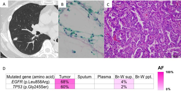
Figure 3. Presentation of case 2.

A. CT findings: A nodule was located in right segment 6; B. Cells showed no atypia on Papanicolaou staining; and C. Histologically, a lepidic pattern of adenocarcinoma was observed on hematoxylin and eosin staining. D. Genomic analyses: Heat map of the mutations detected in each sample. The left column lists the mutated genes with the corresponding amino acid changes. The mutations were detected in both the primary tumor and bronchial wash supernatant. AF, allele fraction; Br-W, bronchial wash; sup., supernatant; ppt., precipitant.
