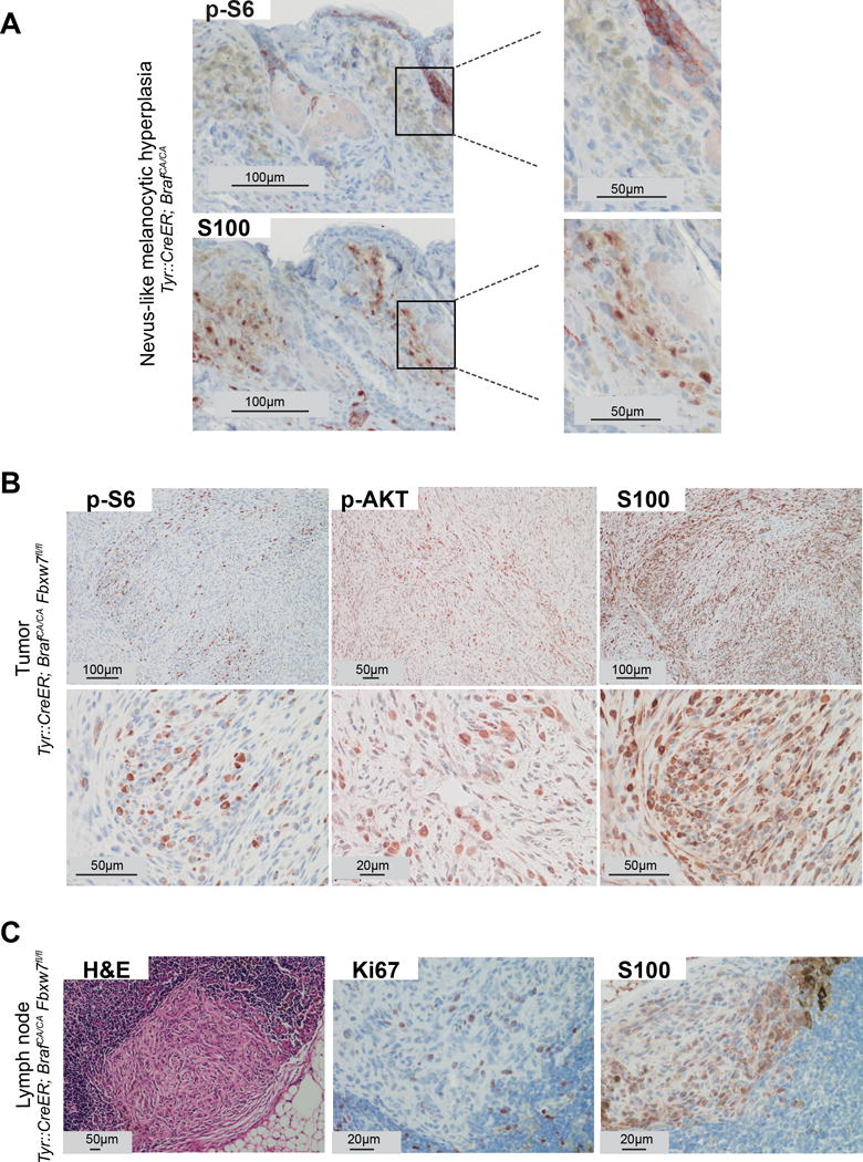
Figure 2.

mTOR signaling is activated during loss of FBXW7-mediated tumorigenesis. (A) Melanocytic hyperplasia (blue nevus-like clones) from Tyr::CreERT2; BrafCA/CA mice stained with p-S6 (upper panel) and S100 (lower panel) using immunohistochemistry. Positive staining for p-S6 within the follicular epithelium is indicated as an internal control. (B) Melanomas (tumors within the deep portion of the dermis) from Tyr::CreERT2; BrafCA/CA; Fbxw7flox/flox mice stained with p-S6, p-AKT (S473), and S100. Low (upper panel) and high power (lower panel) magnifications are shown..(C) Histologic and immunohistochemistry analysis of lymph nodes from Tyr::CreERT2; BrafCA/CA; Fbxw7flox/flox mice stained with H&E, S100, and Ki67. Representative micrographs are indicated.
