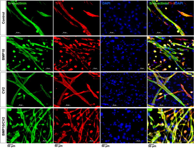
Figure 5. Cardiac marker expression after 14 days of treatment.

Mouse DFAT cells were treated with growth medium supplemented with control vehicle (day 5-7), BMP10 (25 ng/ml, day 5-7), CV2/5-7 (50 ng/ml, day 5-7), BMP10 (day 5-7) followed by CV2 (day 8-10) (BMP10/CV2). Cell morphology and expression of sarcomeric alpha-actinin (Sr-alpha-actinin, green) and Troponin I (Tr-I, red) were examined after 14 days by immunofluorescence. DAPI (blue) was used to visualize nuclei.
