Figure 4.
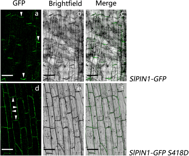
Subcellular localization of the SlPIN1-GFP protein and the phosphomimic SlPIN1-GFP S418D protein. GFP (a,d), bright-field (b,e) and merged (c,f) images were detected using a confocal laser scanning microscopy. (a–c) The basal localization of SlPIN1-GFP on the PM in tomato root cells. (d–f) Nonpolar localization of SlPIN1-GFP S418D on the PM in tomato root cells. The polarity is indicated by the white arrowheads. Scale bar = 50 μm.

