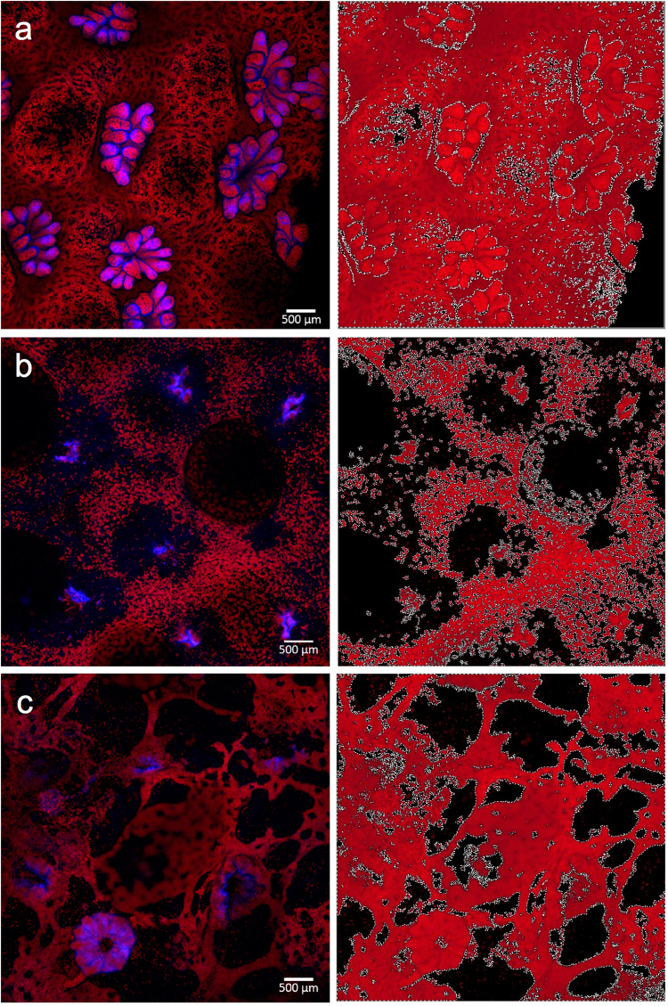
Figure 3.
Image analyses to characterize landscape structure. Confocal microscopy images of (a) healthy, (b) field collected diseased, and (c) laboratory inoculated coral fragments, which were analyzed in Photometrica 7.0 to characterize five measures of landscape structure: total area of fluorescence, edge area, edge to area ratio, number of patches, and patch area. Confocal microscopy images are shown in images on the left, Photometrica analyses of the microscopy images are shown in images on the right. In Photometrica images, the area highlighted in red reflects total area of fluorescence; black regions indicate patches, and white lines indicate edges.

