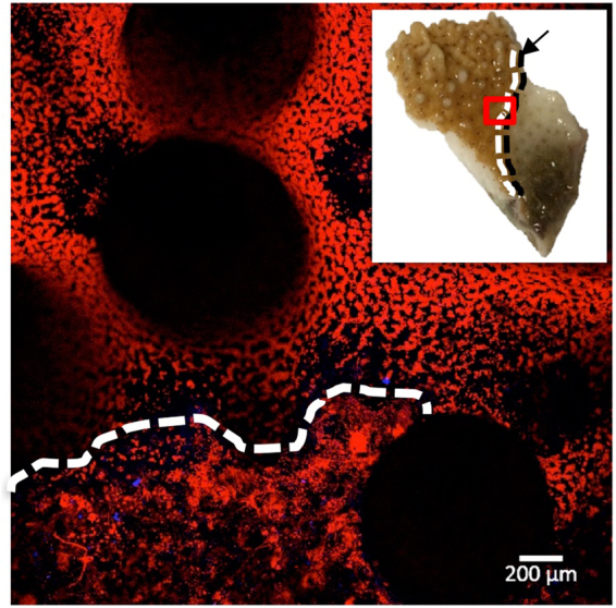
Figure 4.

Linear extension of disease front. Microscopy image of diseased coral fragment. White dotted line indicates the upper limit of fragmented fluorescence adjacent to the disease front. Inset image shows the diseased coral fragment, with a red box indicating the microscope image area, a black arrow and dotted line indicating the visual disease front, and a white dotted line corresponding to the microscopy image of the upper limit of fragmented fluorescence.
