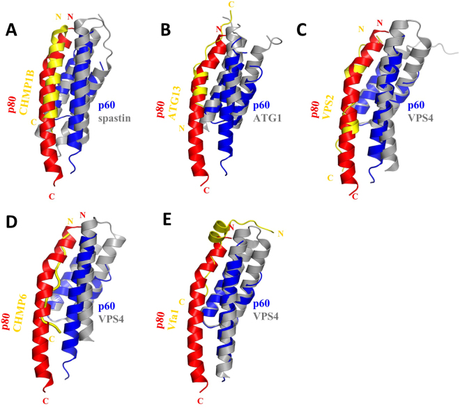
Figure 5.
Comparison of katanin p60-MIT interactions with those of other AAA+ ATPses. (A–E) The structures of different MIT domains are highly similar to each other and they all form an asymmetric three-helix bundle. Superimposition of available complex structures demonstrates an overlap of the binding motifs, although the molecular contacts between these complexes are in most cases not conserved. (A) Spastin-CHMP1B structure (PDB ID 3EAB), (B) ATG1-ATG13 structure (PDB ID 4P1N), (C) Vps4-Vps2 structure (PDB ID 2V6X), (D) Vps4-CHMP6 structure (PDB ID 2K3W), (E) VPS4-Vfa1 structure (PDB ID 5FVK).

