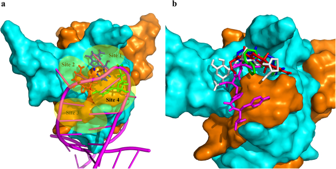
Figure 3.
Potential binding sites on CsrA and docking binding pose of the inhibitors on the CsrA-RNA binding interface. (a) Potential binding sites on the CsrA-RNA binding interface. The interface was formed by the N-terminal of chain A (orange) and Chain B (cyan). RNA was shown as a magenta cartoon and the G10 (red), G11 (blue), A12 (green) of RNA core motif GGA are shown as sticks. (b) The binding pose of inhibitors of CsrA derived from Autodock. Compounds 1 (red), 2 (green), 3 (blue), 4 (magenta) and 5 (white) are shown as a stick.

