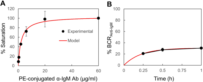
Figure 6.
Determination of the α-IgM Ab concentrations that saturate the surface BCR. (A) Purified spleen B cells were stained with different concentrations of a PE-conjugated α-IgM Ab and analyzed for MFI by FACS. MFI obtained with 60 µg/ml of PE-conjugated α-IgM Ab was set to 100. (B) Proportion of crosslinked BCR in the presence of 1 μg/ml of PE-conjugated α-IgM Ab. Solid circle, actual experimental data; Red line, simulation results. Mean ± SD of 2 independent experiments are shown.

