Fig. 1.
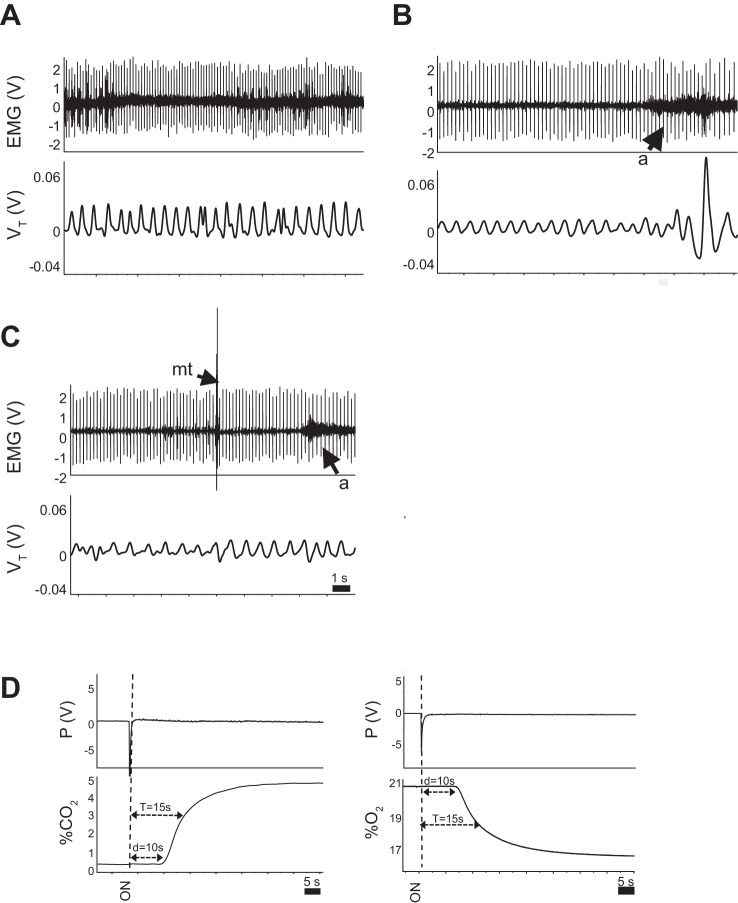
Methodology for assessing sleep state and arousal from sleep. A–C: nuchal EMG activity in a wild-type pup while awake (A), during quiet sleep (QS, B), and during active sleep (AS, C). Note the decrease in amplitude of the EMG in QS, compared with wakefulness, with a further decrease in AS. The appearance of bursts of EMG activity in AS is due to myoclonic twitching (mt), further distinguishing AS from QS. Arousals from sleep (a) are easily discernible on the records by a sustained increase in EMG amplitude. Spikes on the EMG record are the result of cardiac electrical activity, which we used to determine resting heart rate in each sleep state. D: dynamics of gas washin to the chamber are shown for hypercapnia (left) and hypoxia (right). d, delay; T, time constant.

