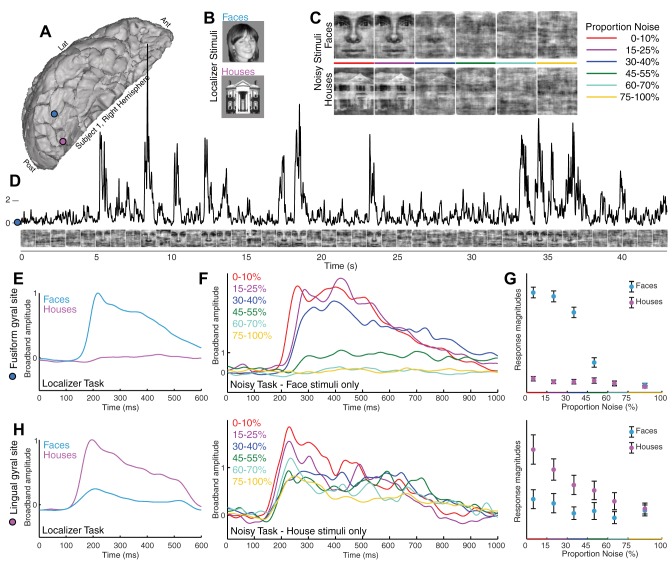
Fig. 1.
Category-selective ventral temporal physiology is graded with stimulus noise. A: ventral temporal ECoG was recorded from fusiform (blue) and lingual (pink) gyral electrodes (subject 1). Ant, anterior; Lat, lateral; Post, posterior. B: localizer task. Simple pictures of whole-field faces and houses were displayed in random order for 400 ms each, with 400-ms blank screen in between. C: noisy task. Phase-scrambled close-up pictures of faces and houses were shown for 1 s each. Stimuli ranged from 0 to 100% noise in 5% increments; subjects pressed a key when they believed a face was shown. D: broadband spectral change in the electrical potential, a reflection of averaged neuronal population activity, is shown above the corresponding stimuli from the noisy task (fusiform site in A). E: averaged broadband response templates to face and house stimuli (from A) are generated from the localizer task. F: averaged broadband responses to face stimuli from the noisy task illustrate diminished and delayed response as image noise increases. G: the face template, generated from the localizer task, is projected to single trials from the noisy task, revealing a robust neurometric function (mean ± SE). At low levels of noise, above the perceptual threshold, there is a parametric decrease in response to faces with increasing noise, with decreasing neuronal activity as image noise is increased. At high noise, above the perceptual threshold, the response drops from a high level to zero response (where all house responses are). H: same as E–G but for the lingual gyral (pink) site in A, with the use of a house template to quantify single-trial responses (far right).

