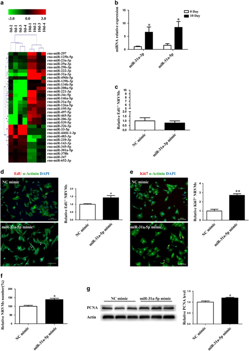
Figure 1.
miR-31a-5p promotes cardiomyocyte proliferation in vitro. (a) MicroRNA (miRNA) arrays identified 32 differentially expressed miRNAs between cardiomyocytes isolated from rats at 0 day and 10 days of age. n=4 per group. (b) Quantitative reverse transcription polymerase chain reaction (qRT-PCR) analysis of miR-31a-3p and miR-31a-5p between cardiomyocytes isolated from rats at 0 day and 10 days of age. n=4 per group. (c) Immunohistochemical stainings for sarcomeric α-actinin and 5-ethynyl-2-deoxyuridine (EdU) staining were determined when neonatal rat ventricular cardiomyocytes (NRVMs) were transfected with the control (NC mimic) or the miR-31a-3p mimic. At least 2000 cells were quantified in each group. (d–f) Immunohistochemical stainings for sarcomeric α-actinin and EdU (d) or Ki-67 (e) followed by quantification of the cell number (f) were performed when NRVMs were transfected with the control (NC mimic) or the miR-31a-5p mimic. At least 2000 cells were quantified in each group. Scale bar: 100 μm. (g) Western blot showed that proliferating cell nuclear antigen (PCNA) expression was increased by the miR-31a-5p mimic. n=3 per group. *P<0.05, **P<0.01 versus the respective control.

