
Figure 3.
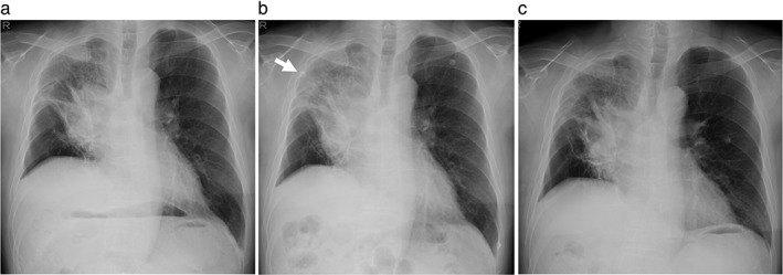
Chest radiograph images during the second nivolumab infusion. (a) Before nivolumab treatment, a lung tumor in the hilar portion of the right upper lobe is observed. (b) One hour after the nivolumab injection, an acute pulmonary infiltrate (arrow) in the upper lung field appeared adjacent to the lung cancer lesion. (c) The pulmonary infiltrate is no longer seen the day after nivolumab administration.