Fig. 3.
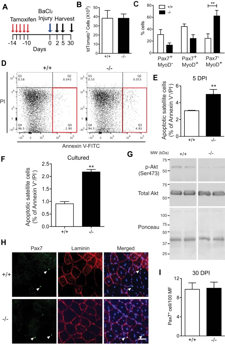
Loss of VCAM-1 alters satellite cell lineage progression and survival but not self-renewal during regeneration. A: schematic of tamoxifen treatment and muscle injury. Gastrocnemius muscles were used for B–G and TA muscles for H and I. B: no difference in the number of tdTomato+ cells 2 days postinjury was observed between the two genotypes. C: tdTomato+ cells were isolated by flow cytometry 2 days postinjury and immediately immunolabeled for Pax7 and MyoD. The percentage of Pax7−MyoD+ cells was significantly increased in Vcam1−/− satellite cells compared with Vcam1+/+. D: representative flow cytometry plots showing increased apoptotic tdTomato+ satellite cells (Annexin V+/PI−) in Vcam1−/− relative to Vcam1+/+ (red boxes). PI, propidium iodide. E: the percentage of apoptotic satellite cells was increased ~1.7-fold in Vcam1−/− relative to Vcam1+/+ muscles. F: apoptotic cells (Annexin V+/PI−) were increased ~2-fold in cultured Vcam1−/− myoblasts relative to Vcam1+/+ myoblasts. G: phospho (p)-AKT levels were decreased in cultured Vcam1−/− myoblasts relative to Vcam1+/+. H: representative images of Pax7 immunolabeling (green) with laminin immunostaining (red) 30 days postinjury. Nuclei were counterstained with DAPI (blue). Scale bar, 50 µm. I: the number of Pax7+ cells/100 myofibers (MF) was not significantly different between genotypes 30 days postinjury (DPI). **P < 0.01; n = 3–5 per genotype.

