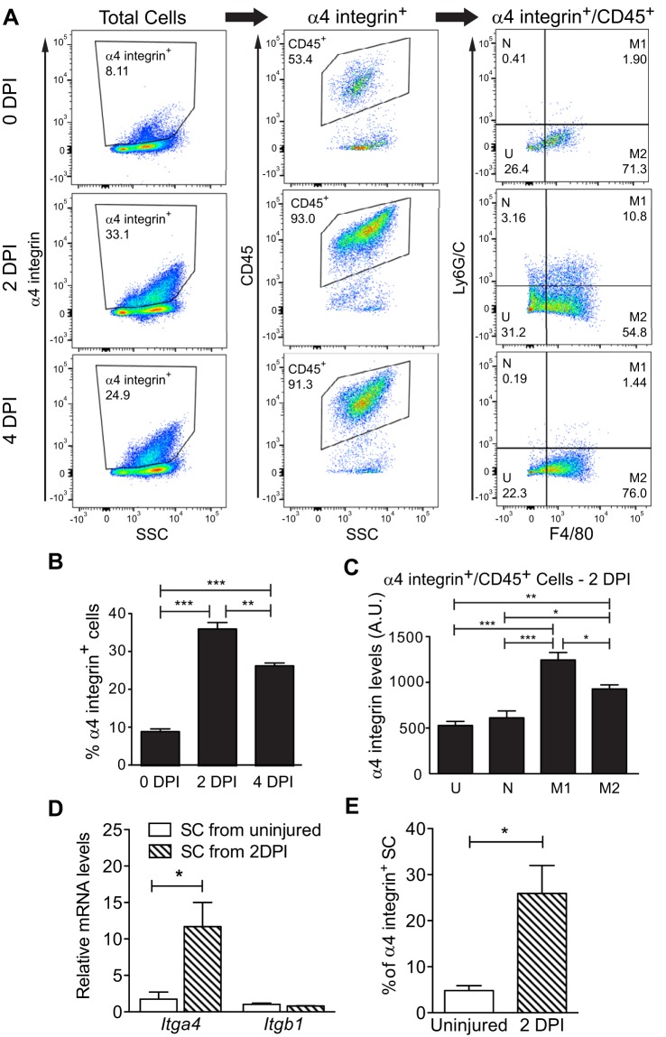
Fig. 4.
Multiple cell types express α4 integrin within regenerating wild-type muscles. A: uninjured hindlimb muscles (0 DPI) or injured gastrocnemius muscles (2 and 4 DPI) were analyzed by flow cytometry for surface levels of α4 integrin, CD45, Ly6G/C, and F4/80. B: the percentage of α4 integrin+ cells increased postinjury. C: M1 macrophages displayed the highest surface levels of α4 integrin compared with other CD45+ cells. DPI, days postinjury; N, neutrophils; M1, M1 macrophages; M2, M2 macrophages; U, uncharacterized CD45+ cells; AU, artificial units; n = 3. D: increased levels of Itga4 (encodes α4 integrin) but not Itgb1 (encodes β1 integrin) mRNA in satellite cells (SC) from injured muscle as detected by RT-qPCR and normalized to Gapdh; n = 3. E: increased percentage of α4 integrin-positive satellite cells after injury as measured by flow cytometry. ***P < 0.001, **P < 0.01, *P < 0.05; n = 4.

