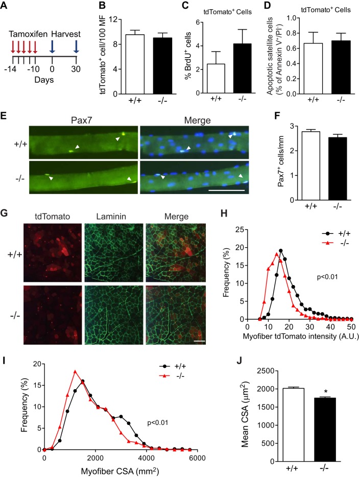
Fig. 5.
Loss of VCAM-1 only alters satellite cell fusion in uninjured muscles. A: schematic of tamoxifen treatment and cell harvest from uninjured muscles. B: the number of tdTomato+ cells per 100 myofibers in sections of tibialis anterior muscles 10 days after the end of tamoxifen treatment did not differ between genotypes. C: by flow cytometry, the percentage of BrdU+ tdTomato+ cells in hindlimb muscles 10 days after the end of tamoxifen treatment did not differ significantly between genotypes. D: by flow cytometry, the percentage of apoptotic tdTomato+ cells in hindlimb muscles 10 days after tamoxifen injection did not differ between genotypes. E: myofibers were isolated from gastrocnemius muscles 10 days after the end of tamoxifen treatment and immunolabeled for Pax7 (green). Pax7+ cells are indicated by arrowheads. Nuclei were counterstained with DAPI (blue). Scale bar, 100 µm. F: no significant difference in the number of Pax7+ cells on isolated myofibers between genotypes; n = 3 mice with >24 myofibers/mouse. G: representative sections of tibialis anterior muscles 10 days after the end of tamoxifen treatment showing tdTomato (red) immunofluorescence and laminin (green) immunostaining. Scale bar, 200 µm. H: histogram depicting frequency distribution of myofiber tdTomato intensity in sections. Vcam1−/− myofibers displayed significantly less tdTomato intensity than Vcam1+/+. AU, artificial units. I: histogram depicting shift to smaller myofiber cross-sectional area (CSA) in Vcam1−/− muscles compared with Vcam1+/+ 40 days after the end of tamoxifen treatment. J: mean myofiber cross-sectional area (CSA) was significantly decreased in Vcam1−/− muscles compared with Vcam1+/+ 40 days after tamoxifen treatment. *P < 0.05.

