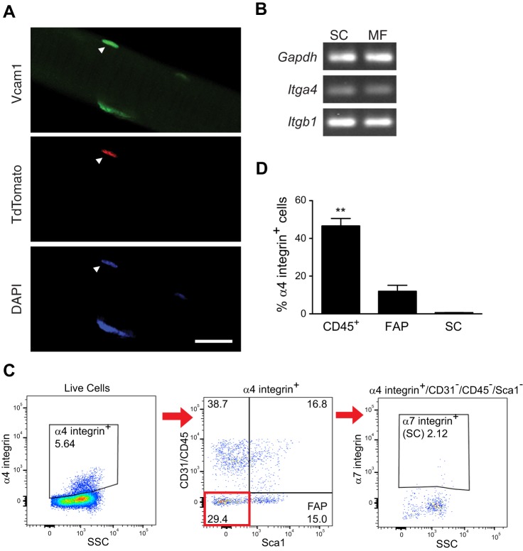
Fig. 6.
Expression of VCAM-1 and its ligands in uninjured wild-type muscles. A: VCAM-1 (green) immunofluorescent labeling of tdTomato+ satellite cell (red) on isolated myofiber. Nuclei were counterstained with DAPI (blue). B: semiquantitative RT-PCR demonstrating that Itga4 (encodes α4 integrin) and Itgb1 (encodes β1 integrin) mRNAs are detected in myofibers. Shown is a representative gel of n = 3 experiments per cell type. C: flow cytometry analysis of α4 integrin+ cells for markers of fibro/adipogenic progenitors (FAP; CD31−/CD45−/Sca1+) and satellite cells (SC; CD31−/CD45−/Sca1−/α7 integrin+). D: the predominant α4 integrin+ mononucleated cell population in uninjured hindlimb muscle was CD45+. **P < 0.01, n = 3.

